![]() This
is a command or new window button. [left mouse] click to execute
This
is a command or new window button. [left mouse] click to execute
![]() This
is a pull down menu. [right mouse] click and hold to see the menu
This
is a pull down menu. [right mouse] click and hold to see the menu
For additional help using any of the software packages please see the SOFTWARE help section of this centers HOMEPAGE.
Written By George Phillips: georgep@rice.edu
Problem1
The purpose of this exercise is to visualize the Ewald sphere construction and the inverse relationship between the size of the reciprocal and direct cells, and to see the effect of moving the crystal on the resulting diffraction pattern. The effect of changes in wavelength on the reciprocal lattice is also illustrated. What you are looking at is a side view of a diffraction experiment, with the X-rays coming in from the left and striking a "crystal" at the center of the sphere. The X-rays are diffracted, resulting in a pattern that appears on the surface of a detector (or piece of film) on the right.
1. Use the pull-down menu labeled "Edit Unit Cell..." under"Options" to set the unit cell parameters to a fairly small cell, 10 x15 x 20 Angstroms. Click on the button labeled "OK", after the cell dimensions are set. Notice that the reciprocal lattice is now relatively large. When the slide bars to the right are moved, notice that as a reciprocal lattice point touches Ewald's sphere the color of the lattice point changes to red and a diffracted ray strikes the detector, producing a "spot" on the detector. Notice that the spacings between spots on the detector is also large, corresponding to the large separation between lattice points on the reciprocal lattice.
2. Now change the unit cell to relatively large values, say 50 x 60 x70 Angstroms. (Be patient, it takes a while to generate a large number of points). Notice that the size of the reciprocal lattice, and the separation between spots on the detector, has shrunk considerably. Note also that the number of spots appearing on the detector has increased. This is a consequence of the smaller reciprocal cell volume. Rotate the crystal with the slide bars. As the crystal is rotated, rings of spots can be seen to come in and out of view. These circles arise from the satisfaction of the von Laue conditions, and represent the intersection of planes in reciprocal space with the Ewald sphere. The diffracted rays around a circle lie on the surface of a cone which is often referred to as a Laue cone.
3. Now change the unit cell dimensions to 10 x 10 x 10 Angstroms, change the wavelength to 2 Angstroms, and change the resolution to 2 Angstroms. Notice now some points are fairly far from the origin of the reciprocal lattice. You can imagine that at even higher resolution (that is, a reciprocal lattice with even greater extent) that some lattice points could never be brought onto the Ewald sphere. This illustrates the concept of the limiting sphere. For a given wavelength, only those lattice points at resolutions greater that 1/2 of the wavelength can ever be observed.
Problem 2
This exercise shows how the observation of Laue circles can be used to precisely align a crystal along a particular axis. Although real crystals can often be aligned approximately by optical examination of the crystal faces, examination in reciprocal space is the best way to achieve an accurate alignment.
1. Set the resolution back to 7 Angstroms and then set the unit cell to about 50 x 60 x 70 Angstroms with a beta angle of 100 degrees. Switch to a simple view of just the diffraction pattern by selecting "View Detector" under the "View" option. Set the axes labeled "Large" and "Phi" to zero. You can use the arrow keys for fine adjustments. Now rotate the crystal around the "Small" axis by putting the pointer on the slide bar and moving it up and down. Note that the pattern rotates, but that the particular spots that are shown do not change. This is a consequence of the circular symmetry of Ewald's sphere about the incident beam and the fact the the "Small" slider rotates the crystal about the incident X-ray beam under these conditions.
2. Now you will orient the crystal so that one of its axes is along the X-ray beam by looking only at the diffraction pattern. use the slide bar labeled "Large" to align the crystal so that its 010 ( or y) axis is along the X-ray beam. (This is a view that will show a non-90 degree angle in the reciprocal lattice. Remember that beta is the angle between the a and c axes and is set to 100 degrees at present. In case you can't find it by wandering around in reciprocal space, the answer is at about +90 degrees around the axis labeled "Large" with "Small" and "Phi" set to zero).
Problem3
The mosaic spread of the crystal (and the degree of monochromaticity of the incident radiation) determine how close a reciprocal lattice point must be to Ewald's sphere for a "reflection" to appear on the detector. The wavelength of the incident radiation also effects the size of the reciprocal lattice.*
1. Use the "Edit Mosaic Spread..." option under "Options" to observe the effect of increasing mosaicity on the resulting diffraction pattern. Note that more spots appear on the detector if the mosaicity, or angular spread of each reciprocal lattice spot, is increased.
2. Now note the effect of a change in wavelength on the pattern by selecting the "Edit wavelength" options under "Options" and clicking on the "Apply" button. (You should click on the gray bar of the wavelength box and drag it out of the way, so you can see the diffraction pattern as you change the wavelength.) Notice that the spots become closer together as the wavelength is decreased and vice versa. Note also that the particular spots that appear on the detector also change, corresponding to the different relative curvature of Ewald's sphere with respect to the lattice.
*Note: An alternative definition is that the size of Ewald's sphere is changed with a change in wavelength. More specifically, some conventions have the radius of Ewald's sphere as 1, with the reciprocal lattice scaled by a multiplicative factor of the wavelength. The other convention has the radius of Ewald's sphere as 1/l, where l is the wavelength with no wavelength dependence on the size of the reciprocal lattice. We have chosen the former so that the relative scaling of data at different wavelengths is preserved for a given crystal to detector distance.
Problem 4
It is possible to use the simulator to integrate the diffraction over some angular range around the Phi axis, resulting in a diffraction pattern that is very much like an oscillation or rotation photograph or a single frame in an area detector data set.
1. This is done by choosing the "Auto Rotation" option to set parameters for the integration. The default parameters call for a 3 degree rotation, with calculations of the spots every 0.2 degree. These will be O.K. for this example. After you click on "OK", then click on the "Start" box in the lower left-hand corner. You can now see more of the reciprocal lattice on the detector, with distinct rows and columns of spots.
The detector can be erased to start another exposure by clicking on the "Clear" box. (The menu at the top of the screen will remain disabled until you click "Clear".)
If one chooses large integration angles (or very large unit cells), some spots overlap with previously recorded ones. This effect can be simulated with this program, and is a real problem that restricts the amount of data that can be collected in one integration.
Problem 5 (optional)
Space groups can be determined (with a few ambiguities) by examination of the reciprocal lattice through X-ray diffraction patterns. By exploring different crystal orientations with the slide bars, determine the likely space groups of some "unknowns" whose parameters have been saved in precreated files. The directions below describe how to load the unknowns. (This is an advanced, difficult exercise, especially if done without peeking at the reciprocal lattice!) If you want to check your answer you can open the "Edit Unit Cell..." option and take a look at the cell constants for the file you chosen as your unknown.
One important piece of information you need here is the geometrical arrangement of the slide bar angles. They are arranged according to the following diagram, which is like a standard goniometer head that is commonly used in the laboratory.
1. Under the "File" option, select "Open", then choose an unknown cell to attempt to determine. Several examples {/usr1/tutorials/} are listed as "unknown1", "unknown2", "unknown3", etc. . It will help if you understand the definitions of the the rotation axes, Small, Large, and Phi (see below). Then you can plan your crystal rotations to examine the diffraction from the crystal in different directions.
3. It will help to take some small, say 3.0 degree oscillation pictures along different directions, as in Exercise 4.
4. If you need a clue, you can sneak a peek at the reciprocal lattice itself by choosing the 'View Side" option.
Small corresponds to the small arc on a goniometer head, Large to the large arc, and Phi is a rotation around the vertical axis. The arrows indicate a positive rotation direction with all angles set to zero in this illustration. Note that because Small is fastened above Large on the goniometer head, as Large increases the Small axis of rotation changes. This is complicated, but is the way real goniometer heads have to be constructed.
      _
     | |
      |
      |
      |
  \_______/      Small is a rotation perpendicular to Large.
      |
 \_________/     Large is a rotation in the plane of the screen.
      |
 ___________     Phi is a rotation about the vertical axis
 |         |
 |_________|
Problem 6
Synchrotrons can provide very intense X-ray beams with a wide range of wavelengths. They can be useful for taking a data set very quickly, sometimes in a millisecond or less. A so-called multi-wavelength "Laue photograph" can be simulated by:
1. Setting the mosaic spread to the minimum value (this reduces "streaking" of the spots on the detector, and illustrates an actual limitation of the Laue method. It is also best to choose unit cell constants in the 40 Angstrom range, with the highest resolution set to 5 Angstroms.
2. The simulation is done by choosing the View Detector option (to save computer time), turning Integration On (also under the View menu header), and then choosing the Edit Wavelength box and systematically decreasing the wavelength from 2.0 to 0.5 in 0.2 Angstrom increments using the small right hand arrow and the pushing the Apply button. (It's tedious, but it works).
Note that circles build up. The reflections at the high resolution limit are no longer the farthest from the center of the pattern, but near the intersections of circles, or nodal reflections.
The above text was writen by George Phillips as part of his XRayView package. To obtain a copy of XRayView email the author: georgep@rice.edu.
This exercise (Exercise1) is designed to familiarize you with the program XtalView, and the process of building residues into an electron density map. A model structure with a poly-alanine region is given in file Exercise1.pdb. The experimental Structure Factors (Fo) obtained from diffraction data are listed in file Exercise1.phs. In this exercise reidues 90 -100 form a poly-alanine chain which you must mutate to the correct sequence. The protein sequence for this crystal is listed in Exercise1.seq. Write down the sequence for residues 90-100, you will use this to create the full model in Xfit.
XtalView is run from a unix shell by issuing the following commands:
sharefonts
xtalmgr &
The Xtalmanager window now appears. You will need to choose or create a project and crystal entry before proceeding. ( The cell dimensions and space group are listed in the file: /usr1/tutorials/exercise1/crystal_Ex1 ).
You may need to create a new directory for Exercise1 and copy the above files from /usr1/tutorials/exercise1 into it. You cannot save your model into the /usr1/tutorials directory, you only have permission to write into your own directories. See the UNIX section below for help.
![]() This
is a command or new window button. [left mouse] click to execute
This
is a command or new window button. [left mouse] click to execute
![]() This
is a pull down menu. [right mouse] click and hold to see the menu
This
is a pull down menu. [right mouse] click and hold to see the menu
 The
PIN in the upper left hand side of a pull down menu will 'stick' the window
open
The
PIN in the upper left hand side of a pull down menu will 'stick' the window
open
After selecting a project and crystal file:
![[Xfit icon]](images/Xfit.gif) Choose
the application XFIT and list the files. Choose
the "*.pdb" and "*.phs" files for exercise1
by clicking on them.
Choose
the application XFIT and list the files. Choose
the "*.pdb" and "*.phs" files for exercise1
by clicking on them.
Click (ADD ARGS) and (RUN). This will bring up the XFIT program with the Exercise1.pdb and Exercise1.phs files loaded.
This first thing that should be done is to calculate the Structure Factors and phases using the model (SFCalc) and from these generate the Electron Density Map usinf a fast fourier transform (FFT).
(SF Calc) --- (Calculate All and Scale)
(FFT)--- Min. resolution 2.0 --- Max. Resolution 8.0 --- (2Fo-Fc) --- (APPLY)
(MODEL) Click on any atom in a residue to add it to the stack ( ALA90 CA ) or type the residue number and hit (Go to).
[Right Mouse] ---> ![]() . . . . . . . . Centers Viewing Area on ATOM on TOP of the Stack
. . . . . . . . Centers Viewing Area on ATOM on TOP of the Stack
[Middle Mouse] . . . . . . . . . . . . . . . . . . . Translate view. Move / rotate / torsion residue {fit mode}
[Left Mouse] . . . . . . . . . . . . . . . . . . . . . . Pick an Atom to go on Top of the Stack
[Left + Middle] . . . . . . . . . . . . . . . . . . . . ZOOM
To Mutate a residue go the the (MODEL) window. Click on the residue you want to mutate, then choose the new residue type that you want. When you have chosen the new residue type, pull down the (Insert Res) menu and choose (Replace and Fit). This fits the new residue into the electron density.
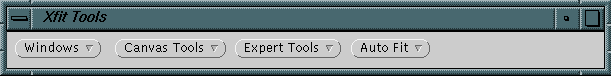
Rotate the model around and check how well the newly placed residue fits the electron density. If the automatic fitting routine does not produce the a good fit then a manual fitting of the residue may be required. The tools for manually fitting a residue are in the pull down menus of the (Xfit Tools) window. Under (Expert Tools) are two other menus (Fit) and (Middle Button Mode).
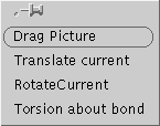 Middle
Button Mode===Moves
residue/residues/parts with middle button
Middle
Button Mode===Moves
residue/residues/parts with middle button
'Translate Current' ======Moves selection
'RotateCurrent' ========Rotates selection around last selection atom picked
'Torsion about bond' ==== Rotates part of selection around bond chosen by picking two atoms
1 2 3 4 5 =========== = Rotates side chain about Chi1 ...
<Space_bar>. . . . . . . . . . . . . . . . . Centers on the next residue.
b . . . . . . . . . . . . . . . . . . . . . . . . . . . Centers on the previous residue.
When you are happy with the placement of your new residue hit <Space_bar> to center on the next residue in the chain and continue mutating the sequence.
After you have finished with all your mutations then save (Output) your model as your_name.pdb. You can also print a picture of your model.
(File) ===== Window for saving, loading PDB files, and loading PHS or SEQ files
(Model) === List the PDB model, edit and modify residues. 'Go To' a specific residue.
(Plot) ===== Set up POSTSCRIPT or RASTER3D publication quality plots.
Follow the instructions for xfit above, only choosing project Exercise2.
In this exercise you will build a sequence of missing residues into an electron density map. The Model, Exercise2.pdb, is complete except for missing residues at 60 - 67. The sequence for these missing residues is given in file Exercise2.seq which will be read into xfit later.
The phase file Exercise2.phs contains the Fobs, Fcalc, and model Phases, so that it can be used to generate an electron density map without using the built-in Fcalc function (SFcalc). So just use the (FFT) window to generate the Electron density from the phase file. The 'Quadratic' spline option produces smoother maps, but will be slower.
The baton command tool simplifies building of an a-carbon backbone into electron density where no previous model exists. The baton commands can be used either from the (Auto Fit) pull down menu, or via the keyboard shortcuts shown.
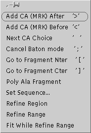
The '>' command adds the next alpha-carbon after the current residue. Once you have more than 5 CA's in a series (fragment) then the 'Poly Ala Fragment' button can be used to create a poly-alanine chain from the CA series. If this fails for any residue then the individual CA (MRK) residue must be manualy mutated to an alanine in the (MODEL) window and then rotated into position using the (Middle Button) commands. When done be sure to hit ';' to exit Baton mode.
Go to the (FILES) window and read in the
sequence file /usr1/tutorials/exercise2/Exercise2.seq
Change the directory to your home (/usr1/tutor01) and then ouput
(save) your model as model.pdb
In the (Baton) window choose 'Set Sequence' to change the sequence of your poly-alanine fragment.
After the sequence is set it will need to be renumbered. In the (MODEL) window choose a residue which has a correct reidue number and then pull down {right_mouse_button} the (SEQUENCE) menu and choose 'Renumber chain'.
Center on the first residue in the mutated fragment and choose (MODEL)(Insert Res)'Replace and Fit'. Hit <space_bar> to center on the next residue and repeat.
When you are finished with model building be sure to SAVE your final model (FILE)'Output'. It is good practise to save your working model at each step before major changes are made. If you do not then your work may be lost.
The final model (model.pdb) is now ready to be refined using a more sophisticated refinement program: XPLOR. (Make sure that the model.pdb file does not contain any 'TER' entries and ends with an 'END')
The refinement parameters have allready been set: so just enter
and watch as the model parameters are refined in real-time. The final output is a table of the cryatllographic R-factor during the refinement. Notice how the R-factor decreases indicating an improvent in the model as refinement proceeds.
The structure determination of a new protein often starts from heavy atom or Multiple Isomorphous Replacement (MIR) maps. The difference between the diffraction from a native crystal and a crystal (or many crystals) soaked in a heavy atom solution(s) is used to find the positions of the heavy atoms in the unit cell. With several heavy atom derivatives combined the 'PHASE PROBLEM' can be solved.
XtalView is run from a unix shell by issuing the following commands:
sharefonts
xtalmgr &
The Xtalmanager window now appears. You will need to choose or create a project and crystal entry before proceeding. ( The cell dimensions and space group are listed in the file: /usr1/tutorials/exercise3/crystal_Ex3 )
![[Xfft icon]](images/Xfft.gif) Choose
the program Xfft and list the files. Choose the Exercise3.df
derivative diffraction data file and the Exercise3.map
ouput file for exercise3. Note that the *.df
file has 4 columns two for each crystal (native and derivative). The two
columns per crystal are for anomalous measurements. If there is no anomalous
signal these two columns are identical.
Choose
the program Xfft and list the files. Choose the Exercise3.df
derivative diffraction data file and the Exercise3.map
ouput file for exercise3. Note that the *.df
file has 4 columns two for each crystal (native and derivative). The two
columns per crystal are for anomalous measurements. If there is no anomalous
signal these two columns are identical.
Click (ADD ARGS) and (RUN). This will bring up the Xffts program.
Check your filenames.
Choose the (Fo*Fo) Patterson (MAP)
option.
Choose your resolution limits ( 12.0 to 4.0 A)
and outlier rejection criterion ( 30% ).
Click on (READ Phase File) and then generate
the map (CALCULATE).
![[Xcontour icon]](images/Xcontour.gif)
Displays a countour map of the PATTERSON difference map generated in Xfft. Remember that for space group 76 P41, the special planes are Z=0.0. 0.25, 0.50, 0.75. A heavy atom peak should be visible on the Z=0.25 plane.
This exercise (exercise5) will demonstrate the crystallographic refinement of a point mutant using the software package XPLOR. The initial model will be that of the native, unmutated, structure. The tyrasine at position 27 has been mutated to a tryptophan. Refinement and Modeling of a Point Mutant In this example we have mutated one point residue Tyrosine 27 to a Tryptophan.
We will start this exercise with the following files: Exercise5.pdb the native structure which is used as a starting model, Exercise5.cell the experimental cell constants and symmetry elements and Exercise5.fob the experimental diffraction intensities We will be using the refinement program XPLOR to refine the atomic positions of all the atoms in the model and the temperature (vibrational/disorder) factors associated with them. The parameter files needed to perform these refinements are ready to be used by XPLOR. The order in which these refinements should be performed is given in the following files:
Please note the required names of all the input, and standard output file names. Always save your model as you proceed, so that in the event of an error you can recover your previous work. Record the 'R' factor after each step of the refinement. Note how the "R" factor decreases as the model is refined against the experimental data, and increases as the resolution is increased.
use the unix command
grep "R=" *.log
to keep track of the R factor as it changes. Examine the model in XtalView's XFIT program ( rigid.pdb and native.pdb)
Examine the Electron Density of the final model at
The following UNIX commands may be usefull.
cp {filenames} {path/filename}...........COPY
mv {filenames} {path/filename}...........MOVE
ls {optional path}.......................LIST files
cd {path}................................Change Directory
cd {../}.................................Back up directory tree
pwd .....................................Print Working Directory
mkdir {dirname}..........................Make a New Directory
more {filenames}.........................Print file to screen
cat {filenames}..........................Print file to screen
man {command}............................Manual for command
help {keyword}...........................List of appropriate commands
grep "string" {filenames}................Search files for string
jot {filename}...........................GUI file editor
Page created by Mark A. White
on or about October 1997. Page last maintained 10/98
Copyright © 1998,
The University of Texas Medical Branch